Screening for PBD-dependent binding inhibitors.

Outlines:

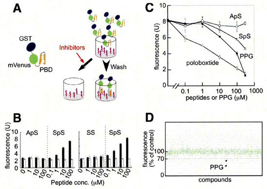
Plk1 possesses a phosphopeptide-binding domain named Polo box domain (PBD) at its C-terminus. PBD is postulated to be essential for Plk1 localization and substrate targeting. We developed a high-throughput screening system to identify inhibitors of PBD-dependent binding and screened a chemical library.
Phosphopeptides (100 uL/well of 10 uM solution, otherwise noted) were covalently bound to maleimide-activated 96-well plates (PIERCE) as instructed by the supplier. GST-mVenus-PBD and its mutant were expressed in E. coli BL21 (DE3). The bacteria were lysed by sonication in sonication buffer (20 mM Tris, pH 8.0/125 mM NaCl/0.5% Nonidet P-40/1 mM EDTA/1 mM DTT/200 uM Na3VO4/50 mM NaF) supplemented with Complete protease inhibitors (Roche Applied Science) and 0.1% lysozyme. After centrifugation, the bacterial lysate was mixed with candidate compounds (at 50 M in the first screening) and put into wells of the phosphopeptide-bound plate and incubated at 4°C overnight. Bound GST-mVenus-PBD was quantitated by spectrofluorometry (ex: 485 nm, em: 530 nm) after five rinses with 0.05% Nonidet P-40 in PBS and two rinses with PBS.


Screening system for inhibitors of PBD-dependent binding and identification of PPG as an inhibitor of PBD-dependent binding.
A. Schematic representation of the screening system. Target phosphopeptides for PBD were covalently bound in 96-well plates, and binding of GST-mVenus-PBD to the phosphorpeptides was quantitated by spectrofluorometry. When inhibitors are present, PBD will fluoresce less. B. Phosphopeptides of PBD-binding sequence (SpS; C-EEEGFGSSpSPVKSPAAP) and their mutated (ApS) or unphosphorylated versions (SS) were bound to 96-well plates using peptides at concentrations as indicated. Binding of wild-type (solid bars) or H538A/K540M mutant forms (open bars) of PBD was determined by spectrofluorometry. C. Effects of various concentrations of phosphopeptides encoding optimal PBD-binding sequence (poloboxtide; MAGPMQSpTPLNGAKK), SpS (C-EEEGFGSSpSPVKSPAAP) and ApS (C-EEEGFGAApSPVKSPAAP) on PBD-dependent binding. Effects of same concentrations of PPG were also examined. In this particular experiment, about 2,000 fluorescence unit of GST-mVenus-PBD were mixed with each competitors, put into each well, washed and bound fluorescence units were plotted. Each symbol represents mean value, while each vertical line indicates standard deviation (n=3). D. Results of initial screening of about 2,500 compounds. The effect of each compound (at 50 μM) on the PBD-dependent binding was standardized and represented as percentage value of control (without compound; ie. 100% means no inhibition). The effects of more than 90% compounds were within the range of 100 ± 20%, only PPG inhibited the binding more than 30%, reproducibly (arrows). (Ref:1)

References:

  1. N. Watanabe, T. Sekine, M. Takagi, J.I. Iwasaki, N. Imamoto, H. Kawasaki,H. Osada.
    Deficiency in chromosome congression by the inhibition of PLK1 polo box domain-dependent recognition.
    J. Bio. Chem., 284, 2344-2353 (2009).

Additional information

Close info