Drug target analysis based on proteomic analysis by 2D-DIGE.

Outline:

The exposure of the cells to bioactive small-molecules induces specific changes of the proteome. Using two-dimensional difference gel electrophoresis (2D-DIGE), proteomic analysis of the HeLa cells treated with a new compound is performed. Based on the expression data of ~300 spots, we compare well-characterized inhibitors and new compounds by hierarchical cluster analysis.

Compounds:Iejimalide A, BNS-22
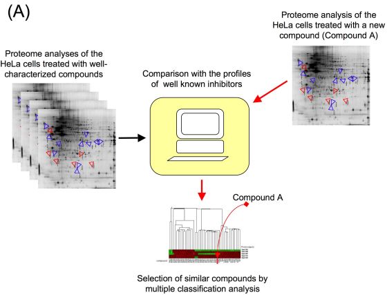


  1. Proteomic profiling system for drug target analysis using 2D-DIGE.(2)
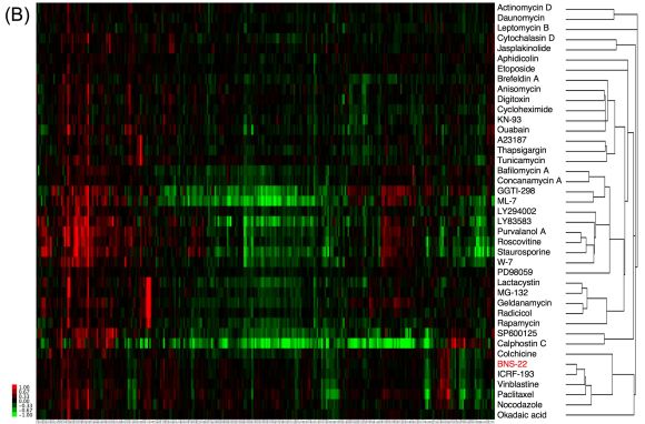
  2. Clustering of BNS-22 and well-known inhibitors based on the data of proteomic analysis using 2D-DIGE.

HeLa cells were treated with compound or with DMSO for 18 h were analyzed by 2D-DIGE and hierarchical cluster analysis was performed using the quantitative data of spots. The target of BNS-22 was estimated to Topo II by the system and inhibitory activity against Topo II was verified. This figure was modified based on the previously published data.(3)

References:

  1. M. Muroi, S. Kazami, K. Noda, H. Kondo, H. Takayama, M. Kawatani, T. Usui, H. Osada.
    Application of proteomic profiling based on 2D-DIGE for classification of compounds according to the mechanism of action,
    Chem. Biol., 17, 460-70 (2010).

  2. K. Wierzba, M. Muroi, H. Osada.
    Proteomics accelerating the identification of the target molecule of bioactive small molecules.
    Curr. Opin. Chem.Biol., 15, 57-65 (2011).

  3. M. Kawatani, H. Takayama, M. Muroi, S. Kimura, T Maekawa, H. Osada.
    Identification of a small-molecule inhibitor of DNA topoisomerase II by proteomic profiling,
    Chem. Biol., 18, 743-751 (2011).

ChemProteoBase

Chemical array screening for small molecule inhibitors.

Outlines:

We have developed a unique photo-cross-linking approach for immobilizing a variety of small molecules in a functional-group-independent manner. Our approach depends on the reactivity of the carbene species generated from trifluoromethyldazirine upon UV irradiation. It was demonstrated in model experiments that the photogenerated carbenes were able to react with every small molecule tested, and they produced multiple conjugates in most cases. It was also found in on-array immobilization experiments that various small molecules were immobilized, and the immobilized small molecules retained their ability to interact with their binding proteins. With this approach, photo-cross linked microarrays of 2000 natural products and drugs were constructed. This photo-cross-linked microarray format was found to be useful not merely for ligand screening but also to study the structure-activity relationship, that is, the relationship between the structural motif (or pharmacophore) found in small molecules and its binding affinity toward a protein, by taking advantage of the nonselective nature of the photo-cross-linking process.
その他:化合物の数が1000以上まとまっていれば、単独で化合物アレイを作製することが可能である。化合物の数が少ない場合は、化合物が集まった段階で化合物アレイを作製するため、作製までに時間を必要とする。


Overview of Systematic Chemical Array Screening Method.
Human genes of interest are cloned and restriction enzyme-digested fragments are inserted into pDsRed-express and sequenced. GLORIA contains 100 genes, which can be expressed as RFP-fused proteins in mammalian cells. HEK 293T cells are transfected with vectors encoding RFP fused-proteins or RFP for 24 h. After transfection, cell lysates are prepared for incubation with glass slides, where compound from NPDepo are immobilized in duplicate by a photo-crosslinking technique. Detection of "hit ligands" is done by merged display analysis. Spots that interact with RFP or RFP-fused proteins are designated green and red respectively. After two slides are merged, true hit compounds are detected as red signals, while false positive signals are yellow.(Ref: 1)

References:

  1. I. Miyazaki, S. Simizu, H. Ichimiya, M. Kawatani, H. Osada.
    Robust and systematic drug screening method using chemical arrays and protein library: Identification of novel inhibitors of carbonic anhydrase II.
    Biosci. Biotechnol. Biochem., 72, 2739-2749 (2008).

  2. N. Kanoh, A. Asami, M. Kawatani, K. Homda, S. Kumashiro, H. Takayama, S. Shimizu, T. Amemiya, Y. Kondoh, S. Hatakeyama, K. Tsuganezawa, R. Utata, A. Tanaka, S. Yokoyama, H. Tashiro, H. Osada.
    Photo-cross-linked small-molecule microarrays as chemical genomic tools for dissecting protein-ligand interactions.
    Chem.Asian J., 1, 789-797 (2006).

  3. N. Kanoh, S. Kumashiro, S. Simizu, Y. Kondoh, S. Hatakeyama, H. Tashiro, H. Osada.
    Immobilization of natural products on glass slides by using a photoaffinity reaction and the detection of protein-small-molecule interactions.
    Angew. Chem. Int. Ed., 42, 5584-5587 (2003).

Identification of target proteins using small molecule affinity matrix.

Outlines:

Affinity purification of small-molecule binding protein has been one of the most important techniques in forward chemical genetics. Once hit compound(s) which induce a specific phenotype in a cellular system are identified, the next step is to identify the molecular target.
Recently, we reported a coupling method that enables the introduction of a variety of small molecules onto glass slides through a photoaffinity reaction. By using this method, aryldiazirine groups covalently attached to glass slides are transformed upon irradiation with UV light into highly reactive carbenes, which in turn bind to or insert irreversibly into a proximal small molecule in a manner that is independent of the functional groups. This method, referred to as photo-cross-linking, has proven useful in the construction of small-molecule microarrays. It is expected that a similar chemical approach could be used to introduce small molecules onto affinity gel, such as onto agarose beads.
We have prepared PALC beads and demonstrated that photo-cross-linked small-molecule agarose beads can be used to identify and purify the binding protein. It was of note that complex small molecules such as FK506, rapamycin, and cyclosporin A could be introduced on the affinity matrix by using a uniform and straightforward procedure.


Identification of M-GFN-binding proteins.
(A) Model structure of M-GFN beads. (B) Detection of the coprecipitated proteins for M-GFN beads from RAW264 cell lysates. RAW264 cell lysates were precleared with control beads and incubated with M-GFN beads. The reacted beads were washed, and the eluted proteins were subjected to SDS/PAGE and visualized by CBB staining. The coprecipitated proteins for M-GFN beads were identified as described in Materials and Methods. (C-E) Purified His-tagged SGTA (C), GLO1 (D), or SCP2 (E) protein was incubated with control beads and M-GFN beads in the presence or absence of M-GFN as a competitor. The reacted beads were washed, and the eluted proteins were immunoblotted with anti-Xpress Ab, which allows detection of recombinant His-tagged proteins containing the N-terminal leader peptide (Xpress epitope). C, Control beads; M, M-GFN beads.(Ref:2)

References:

  1. N. Kanoh, K. Honda, S. Simizu, M. Muroi, H. Osada.
    Photo-crosslinked small molecule affinity matrix for facilitating forward and reverse chemical genetics.
    Angew. Chem. Int. Ed., 44, 3559-3562 (2005).

  2. M. Kawatani, H. Okumura, K. Honda, N. Kanoh, M. Muroi, N. Dohmae, M. Takami, M. Kitagawa, Y. Futamura, M. Imoto, H. Osada.
    The Identification of an osteoclastogenesis inhibitor through the inhibition of glyoxalase I.
    Proc. Natl. Acad. Sci. U S A., 105, 10691-11696 (2008).

SPR imaging of photo-cross-linked small molecule microarrays on gold.

Outlines:

We have recently developed a "photoaffinity" surface plasmon resonance (SPR) imaging platform to enable the immobilization of structurally diverse small molecules on solid supports in a "functional group-independent" manner.
A gold-coated glass chip (TOYOBO, Japan) was immersed in the ethanol solution, which contained 0.1 mM photo-cross-linker and 0.9 mM dummy linker, for 16 h. The chip was rinsed successively with ethanol, water, and ethanol and dried under an air stream to obtain a photoreactive chip. DMSO solutions (10 mM) of small molecules were spotted on the chip using an automated spotter (TOYOBO, Japan). The obtained chip was dried in vacuo to evaporate the DMSO, and the chip was then irradiated under a UV transmission filter (Sigma-Koki, Japan) with a Stratalinker 1800 UV cross-linker (Stratagene, CA) or a CL-1000L ultraviolet cross-linker (UVP Inc., CA). The irradiated energy on the chip amounted to 2.8 J/cm2. The surface was repeatedly rinsed with DMSO, water, and ethanol.
The photo-cross-linked small-molecule array was placed in an SPR imaging instrument (TOYOBO, Japan). The SPR signals were obtained in the running buffer (10 mM HEPES, 150 mM NaCl, pH 7.2). The running buffer and the protein samples in the same buffer were applied to the array surface at 100 uL/min. All SPR experiments were performed at 30 oC. The SPR image and signal data were collected with an SPR analysis program (TOYOBO, Japan). The SPR difference image was constructed by using Scion Image (Scion, MD).


Overview of small-molecule microarrays.
a) Upon printing, a library of small molecules with functional group A readily attach to the solid surface that is derivatized with functional group B, which is reactive to A. Coupling between an inadequate pair of functional groups (i.e., B and C) does not occur. b) When the small molecules are properly immobilized through a selective coupling (D), interactions between these molecules and their binding proteins can be detected sensitively. However, when the immobilization is inadequate (e.g., E), the immobilized small molecule is useless for the protein binding assay. Nonselective immobilization (F) enables not only the introduction of a variety of small molecules, but also the production of a variety of conjugates, some of which are expected to retain affinity toward the binding protein. c) The ideal functional group (G) should be activated under mild conditions, and the resulting species should immobilize a variety of small molecules in a functional-group-independent manner. Moreover, the functional group should not remain on the surface after the immobilization process.(Ref:3)

References:

  1. K. Kawai, A. Saito, T. Sudo, H. Osada.
    Specific Regulation of Cytokine-Dependent p38 MAP Kinase Activation by p625/SQSTM1.
    J. Biochem., 143, 763-772 (2008).

  2. A. Saito, K. Kawai, H. Takayama, T. Sudo, H. Osada.
    Improvement of photoaffinity SPR imaging platform and determination of the binding site of p62/SQSTM1 to p38 MAP kinase.
    Chem. Asian J., 3, 1607-1612 (2008).

  3. N. Kanoh, M. Kyo, K. Inamori, A. Ando, A. Asami, A. Nakao, H. Osada.
    SPR imaging of photo-cross-linked small molecule microarrays on gold.
    Annal. Chem., 78, 2226-2230 (2006).

Screening for PBD-dependent binding inhibitors.

Outlines:

Plk1 possesses a phosphopeptide-binding domain named Polo box domain (PBD) at its C-terminus. PBD is postulated to be essential for Plk1 localization and substrate targeting. We developed a high-throughput screening system to identify inhibitors of PBD-dependent binding and screened a chemical library.
Phosphopeptides (100 uL/well of 10 uM solution, otherwise noted) were covalently bound to maleimide-activated 96-well plates (PIERCE) as instructed by the supplier. GST-mVenus-PBD and its mutant were expressed in E. coli BL21 (DE3). The bacteria were lysed by sonication in sonication buffer (20 mM Tris, pH 8.0/125 mM NaCl/0.5% Nonidet P-40/1 mM EDTA/1 mM DTT/200 uM Na3VO4/50 mM NaF) supplemented with Complete protease inhibitors (Roche Applied Science) and 0.1% lysozyme. After centrifugation, the bacterial lysate was mixed with candidate compounds (at 50 M in the first screening) and put into wells of the phosphopeptide-bound plate and incubated at 4°C overnight. Bound GST-mVenus-PBD was quantitated by spectrofluorometry (ex: 485 nm, em: 530 nm) after five rinses with 0.05% Nonidet P-40 in PBS and two rinses with PBS.


Screening system for inhibitors of PBD-dependent binding and identification of PPG as an inhibitor of PBD-dependent binding.
A. Schematic representation of the screening system. Target phosphopeptides for PBD were covalently bound in 96-well plates, and binding of GST-mVenus-PBD to the phosphorpeptides was quantitated by spectrofluorometry. When inhibitors are present, PBD will fluoresce less. B. Phosphopeptides of PBD-binding sequence (SpS; C-EEEGFGSSpSPVKSPAAP) and their mutated (ApS) or unphosphorylated versions (SS) were bound to 96-well plates using peptides at concentrations as indicated. Binding of wild-type (solid bars) or H538A/K540M mutant forms (open bars) of PBD was determined by spectrofluorometry. C. Effects of various concentrations of phosphopeptides encoding optimal PBD-binding sequence (poloboxtide; MAGPMQSpTPLNGAKK), SpS (C-EEEGFGSSpSPVKSPAAP) and ApS (C-EEEGFGAApSPVKSPAAP) on PBD-dependent binding. Effects of same concentrations of PPG were also examined. In this particular experiment, about 2,000 fluorescence unit of GST-mVenus-PBD were mixed with each competitors, put into each well, washed and bound fluorescence units were plotted. Each symbol represents mean value, while each vertical line indicates standard deviation (n=3). D. Results of initial screening of about 2,500 compounds. The effect of each compound (at 50 μM) on the PBD-dependent binding was standardized and represented as percentage value of control (without compound; ie. 100% means no inhibition). The effects of more than 90% compounds were within the range of 100 ± 20%, only PPG inhibited the binding more than 30%, reproducibly (arrows). (Ref:1)

References:

  1. N. Watanabe, T. Sekine, M. Takagi, J.I. Iwasaki, N. Imamoto, H. Kawasaki,H. Osada.
    Deficiency in chromosome congression by the inhibition of PLK1 polo box domain-dependent recognition.
    J. Bio. Chem., 284, 2344-2353 (2009).

Screening for tyrosine phosphatase inhibitors.

Outlines:

Reversible phosphorylation on tyrosine residues of proteins is one of the earliest and important events in the signal transduction pathways leading to the stimulation of cell proliferation. Because many oncogenes encode protein tyrosine kinases (PTKs), much attention had been paid to PTK compared with protein tyrosine phosphatases (PTPases). Recently, it was reported that PTPase played an important role in the signal transduction. Mitosis is initiated following the activation of cdc2-cyclin B kinase complex, so-called M-phase promoting factor or maturation promoting factor (MPF). The key step in the activation of MPF is dephosphorylation on Thr-14 and Tyr-15 of cdc2 protein by cdc25, a kind of dual-specificity protein phosphatases. VHR (VHl-related human protein), which was isolated from a human fibroblast has the dual-specificity phosphatase activity towards phosphotyrosine and phosphoserine/threonine like cdc25. The receptor-type PTPase, CD45 (a leukocyte common antigen) activates protein tyrosine kinase p56 Ick and p59 fyn , which are associated with the intracellular domain of the T cell surface glycoproteins, CD4 and CD8. CD45 is involved in T cell and B cell development and activation. Specific PTPase inhibitors should be useful as a biological tool to reveal the signal transduction. Okadaic acid and tautomycin, which are known to inhibit serine/threonine protein phosphatases (PPases), are valuable tools to test the physiological role of serine/threonine phosphorylation in the signal transduction. On the contrary, only a few PTPase inhibitors, for example, sodium orthovanadate (vanadate), phenylarsine oxide (PAO) and dephostatin have been known. A more potent and selective PTPase inhibitor is still required. To discover specific PTPase inhibitors, we have started the screening of microbial metabolites.
GST-VHR fusion protein (VHR) and GST-cdc25B fusion protein (cdc25B) was expressed in bacteria. Dephosphorylation activity of VHR and cdc25B to p-nitrophenylphosphate (pNpp) was measured in the assay buffer (25 mM MOPS, pH 6.5, 5 mM EDTA, 1 mM dithiothreitol).


The lineweaver-Burk plot of RK-682 (A) and vanadate (B) to pNPP.
The concentrations of RK-682 were 2.7 (●), 4.1 (○), 5.4 (■)uM. VHR (20 ug/ml) hydrolyzed pNPP (1, 2, 3, 4, 5 mM). Vanadate concentrations were 0 (●), 0.16 (○), 0.25 (■)mM. (Ref:2)

References:

  1. T. Hamaguchi, A. Takahashi, A. Manaka, M. Sato, H. Osada
    TU-572, a potent and selective CD45 inhibitor, suppresses IgE-mediated anaphylaxis and murine contact hypersensitivity reactions.
    Int. Arch. Allergy Immunol., 126, 318-324 (2001).

  2. T. Hamaguchi, T. Sudo, H. Osada.
    RK-682, a potent inhibitor of tyrosine phosphatase, arrested the mammalian cell cycle progression at G1 phase.
    FEBS Lett., 372, 54-58 (1995).

Inhibition of PDI activity.

Outlines:

 Almost secreted proteins will involve in disulfide bond conformation which occur intracellular leading to the activated protein. Moreover, cancer metastasis-related proteins and growth factors are secreted. Therefore, unknown cancer metastasis factors and its functional mechanisms can be evaluated with PDI inhibitor. Beside this, compounds showing the inhibitory activity can be evaluated on cancer metastatic effect with outside institution collaboration is possible.
その他:阻害活性が見出された物質はがん転移阻害活性評価を外部機関に依頼することも可能。

Screening for heparanase inhibitors.

Outlines:

Heparanase is an endo-beta-D-glucuronidase and plays important roles in tumor metastasis. When tumor cells spread out, they must penetrate the extracellular matrix (ECM) and the basement membrane (BM). At that time, tumor cells secrete matrix metalloproteases and heparanase to degrade major components of ECM and BM such as type IV collagen and heparan sulfate proteoglycans. Moreover, heparanase releases several growth factors, such as bFGF and VEGF, which bind toeparan sulfate (HS) chains in ECM and BM tocause angiogenesis. Therefore, heparanase is recognized as a target molecule for antitumor agents.
Heparan sulfate (HS) was labeled with N-(4-nitro-2,1,3,-benzoxadiazoyl)-N-methyl-2-aminoacetohydrazide (NBD-CO-Hz). The cells were washed suspended in PBS (pH 6.0)., and disrupted by 5 cycles of freezing and thawing. The lysates were centrifuged at 15,000 rpm for 15 min at 4 oC. A mixture of 27 ul of cell lysate and 3 ul of fluorescence-labeled HS solution (0.1 ug/ul in PBS, pH 6.0) was incubated at 37 oC for 24 h. Following the addition of 6 ul of sampling solution (36% glycerol, 1% bromophenol blue), 20 ul of each reaction mixture was subjected to SDS-PAGE (20%), and the gel was scanned by the Variable Mode Imager Typhoon 9400 (Amersham Biosciences).
その他:阻害活性が見出された物質はがん転移阻害活性評価を外部機関に依頼することも可能。

Consequence of the transfection of heparanase gene (A) and substrate specificity of the HpeG2-HP-1 lysate.
(A) haprana sulfate (HS) was treated with the lysate of the indicated cells at 37 oC for 24 hours. (B) Glycosaminoglycans were incubated with or without the lysate at 37 oC for 24 hours. HS: haparan sulfate, HR: heparin, CS: chondroitin sulfate, KS: keratan sulfate. (Ref:2)

References:

  1. N. S. Lai, S. Simizu, D. Morisaki, M. Muroi, H. Osada.
    Requirement of the conserved, hydrophobic C-terminus region for the activation of heparanase.
    Exp. Cell. Res., 314, 2834-2845 (2008).

  2. K. Ishida, T. Teruya, S. Simizu, H. Osada.
    Exploitation of Heparanase Inhibitors from Microbial Metabolites Using an Efficient Visual Screening System.
    J. Antibiot., 57, 136-142 (2004).

Screening for inhibitors or activators of MAP kinases.

Outlines:

The cellular response to diverse external stimuli is controlled via a complex array of phosphorylation cascades. The ERK cascade is a prominent component of the MAP kinase family that, in particular, plays integral role in both growth factor and stress signalling. Recent studies identified members of the MAPK superfamily that include JNK/SAPK and p38 MAPK, which are activated by various environmental stresses such as UV irradiation or exposure to agents, in addition to classical MAPKs. Each MAPK group has a distinct substrate specificity and is regulated by a separate signal transduction pathway. Mammalian cells, therefore, contain multiple MAPK superfamily signal transduction pathways that mediate the effects of extracellular stimuli on a wide array of biological processes. The activities of these various MAPKs can be measured directly in acrylamide gel containing various substrates.
Cells (1 x 10⁶) treated/untreated with various drugs were lysed in lysis buffer and the protein concentration was determined. To assay total cell lysate, equal amounts of protein (50-100 ug) were electrophoresed in 10 % SDS-PAGE containing either MBP (0.5 mg/ml), GST-c-Jun (1-79; 0.1 mg/ml), or GST-ATF2 (0.1 mg/ml) as a substrate. After electrophoresis, SDS was removed from the gel, protein was renatured, and a kinase assay was carried out by incubating the gel in buffer containing [gamma-32P]ATP. Gels were washed and dried, and the incorporated radio activity was analyzed by autoradiography.


Cylolrienin A activates a kinase with an apparent molecular mass of 36 kDa with kinetics different from JNK/SAPK and p38 MAPK in HL-60 cells.
(A) in-gel kinase activity of cells treated with cytotrienin A. Cells were incubated with cytotrienin A at the concentrations indicated. Cell lysates were eleclrophoresed in 10% SDS-PAGE, and kinase activity was determined by an in-gel kinase assay using MBP as a substrate. (B) kinetics of the activation of p.36 MBP kinase. JNK/SAPK. and p.38 MAPK during apoptosis induced by cytotrienin A. Cells were incubated with 100 ng/ml cytotrienin A for the times indicated, and the activation of the JNK/SAPK and p38 MAPK was analyzed by Western blotting using antibodies that specifically recognize the activated JNK/SAPK and p38 MAPK kinases. The p36 MBP kinase activity was measured as described in A. (C) effect of a specific inhibitor of p38 MAPK, SB203580, on p36 MBP kinase activation. Cells were treated with 100 ng/ml cytotrienin A in the presence of SB203580. After incubating for 2 h, p38 MAPK activation and p36 MBP kinase activity were measured as described in B. (Ref:1)

References:

  1. H. Kakeya, R. Onose, H. Osada.
    Caspase-mediated activation of a 36-kDa myelin basic protein kinase during anticancer drug-induced apoptosis.
    Cancer Res., 58, 4888- 4894 (1998).

Screening of novel antifungal agents through the morphological change in rice blast fungi.

Outlines:

Upon exposure to one drug, rice blast fungus Magnaporthe oryzae often dynamically and specifically changes their shape depending on the mode of action of the drug. It prompted us to construct the encyclopedia of fungus cell morphology, consisting of the unique cell-shape changes induced by various compounds, including well-defined antifungal agents. Particularly, we have examined the effect of 200 routinely used compounds in a time- and dose-dependent manner and investigated the relationship between the phenotypes and the action mechanism of drugs. Now, we could easily discriminate the phenotypes induced by well-known antifungals, such as polyoxins (chitin synthesis; swelling), azoles (ergosterol synthesis; toxic) and griseofulvin (tubulin; short). Our screening system allows us to classify test compounds to classes of compounds sharing similar mechanism of action and to identify antifungal compounds with novel mechanism of action effectively.

作用既知薬剤:Polyoxin A

control Polyoxin A
Polyoxin A, a chitin synthesis inhibitor, induces characteristic swelling in M.oryzae.

More Articles ...

  1. p38 MAP kinase inhibitor screening using transformed bacteria.
  2. Screening for HIV-1 Vpr inhibitors.
  3. The bleb forming assay for the activators and inhibitors of PKC.
  4. Screening for compounds with morphological reversion activity on transformed cells.

Additional information

Close info