
")
Screening of cell cycle inhibitors by a bioassay using a mouse cdc2 mutant cell line, tsFT210.
Outlines:
The analysis of tsFT210, a mouse temperature-sensitive mutant with a defect in the cdc2 gene, demonstrated that the cdc2 kinase is essential for triggering mitosis. Similarly, there are also many reports which describe the application of inhibitors in elucidating the regulatory mechanism of the cell cycle. Among them, it was shown that a protein kinase inhibitor, staurosporine, inhibits the cell cycle progression at G2 phase through the inhibition of cdc2 kinase. Based on these recent findings, we have established a simple and rapid system utilizing the temperature sensitive mutant, tsFT210cells, to detect cell cycle inhibitors.
A temperature sensitive mutant, tsFT210, which was isolated from the mouse mammary carcinoma cell line FM3A, was maintained in suspension culture in RPMI-1640 medium supplemented 5% calf serum at 32 oC. The tsFT210 cells were synchronized at the G2 phase by culture at 39.4 oC (restrictive temperature) for 17 h.
In the asynchronous-culture assay, cells cultured at the permissive temperature of 32 oC were seeded into a 12-well plate at a density of 2 x 10⁵ cells/ml in 1 ml of fresh medium. Then cells were treated with inhibitors and cultured for 17 h at 32 oC.
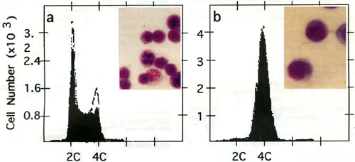
In the synchronous-culture assay, cells were seeded at a density of 2 x 10⁵ cells/ml into a 12 well plate and were pre-incubated at 39.4 oC for 17 h to obtain G2-arrested cells. Then, 10 ul of each sample solution was added, and the cells were incubated at 32 oC for 4 h. After incubation, morphological characteristics of the cells were examined directly by microscopic observation. The cells were also subjected to flow cytometric analysis to confirm the DNA contents in cells.
Examples of positive compounds:
Pironetin, Terpendole E, Cytochalasins, Paclitaxel, Vinblastine
Effects of sangivamycin, RK-1409 and RK-286C on the cell cycle progression from G2- to G1-phase. Asynchronized tsFT210 cells cultured at (a) 32 oC were synchronized at the G2 boundary by incubation at (b) 39 oC for 17 h. The cells were then shifted to 32 oC to release them from G2 arrest. The cells passed through M phase and entered G1 (c) after 4 h. When the cells were simultaneously released from the temperature-arrest in the presence of (d) 10 uM sangivamycin, (e) 0.3 uM RK-1409 and (f) 1.6 uM RK-286C, the cell cycle progression from G2- to M-phase was inhibited. Photographs show morphological characteristics of the corresponding cells observed under the microscope. (Ref:1)
References:
H. Osada, C.-B. Cui, R. Onose, F. Hanaoka.
Screening of cell cycle inhibitors from microbial metabolites by a bioassay using a mouse cdc2 mutant cell line, tsFT210.
Bioorg. Med. Chem., 5, 193-203 (1997).